Users report:
Comparison of Images Obtained by DEI and Refraction-Contrast Imaging. by N.Yagi, R.Lewis & C.Hall (PDF in English)
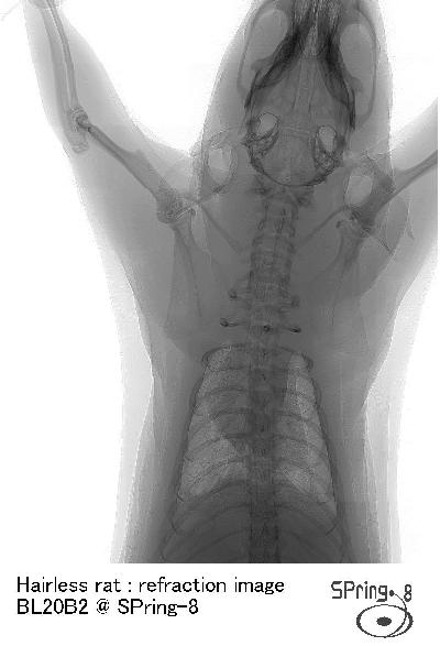
Refraction-enhanced imaging experiments at BL20B2
A hairless rat was imaged. This image was reconstructed from scanned images. The images were recorded with a "Beam Monitor 4" with a C4880-75 cooled CCD camera.
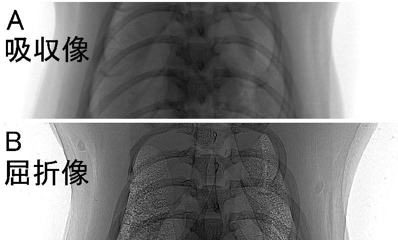
This is a comparison of images with short and long distance between the rat and the detector. "A" is with a short distance (absorption image), "B" is with a long distance (refraction image).
Reference:
N.Yagi, Y.Suzuki, K.Umetani, Y.Kohmura and K.Yamasaki. "Refraction-enhanced x-ray imaging of mouse lung using synchrotron radiation source." Medical Physics 26(10), 2190-2193 (1999)
Y. Suzuki, N. Yagi, and K. Uesugi. "X-ray Refraction-enhanced Imaging and a Method for Phase-retrieval for a Simple Object." J. Synchrotron Rad. 9, 160-165 (2002)
M. J. Kitchen, D. Paganin, R. A. Lewis, N. Yagi, K. Uesugi and S. T. Mudie. "On the Origin of Speckle in X-ray Phase Contrast Images of Lung Tissue." Phys. Med. Biol. 49, 4335-4348 (2004)