These are collection of monochromatic CT images obtained at BL20B2.
Detectors were Beam Monitor 1, 2 or 4.
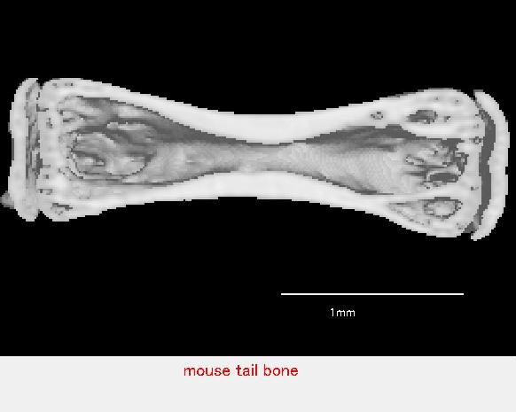
Tail of a small mouse.
The contrast was adjusted to see the bone, and a section was cut.
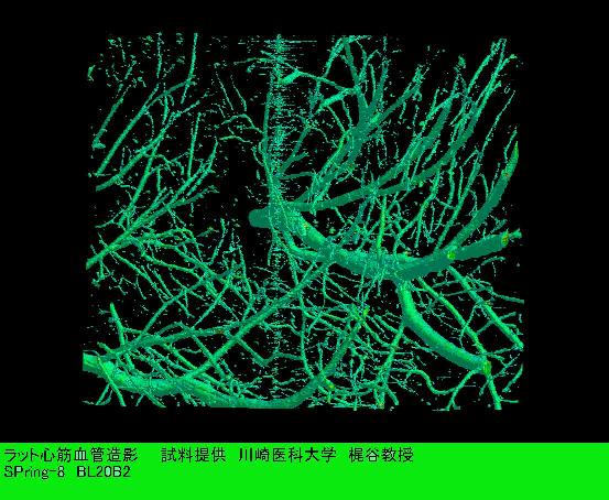
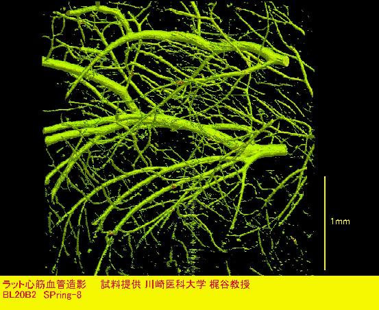
Rat heart
Blood vessels in the wall of heart was filled with a contrast agent (Ba).
Volume rendering was done on the CT image.
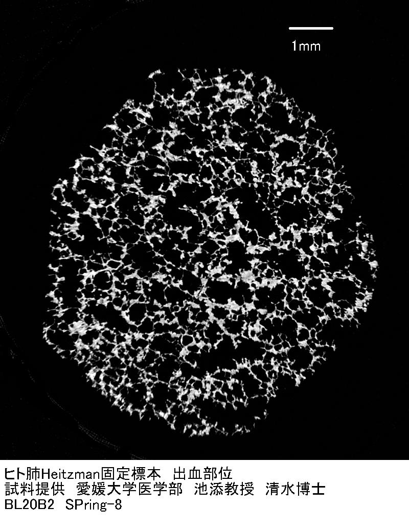
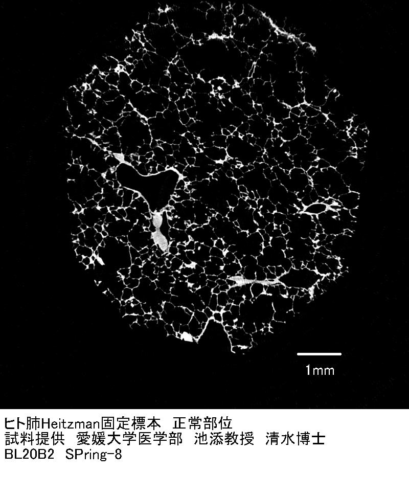
Heitzman preparation of human lung. Normal area is shown.
This is an area of lesion.